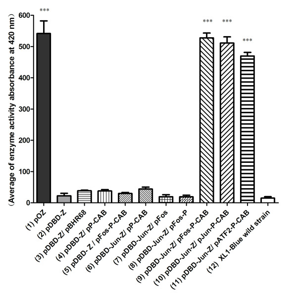
Figure 2.
β-Galactosidase activity assay. E. coli XL1-Blue harboring relative plasmids were cultured in LB medium with 20 g/L glucose for 14 h at 37°C, 200 rpm, then β-galactosidase activity was assayed by Bacterial X-Gal Staining Kit (GENMED, Shanghai, China). Bar 1: LacZ has a strong expression under control of phaP promoter (positive control); Bar 2: LacZ expression can be repressed by DBD; Bar 3-5: PHB granules, PhaP, and bFos-PhaP can not pull down DBD from phaP promoter to release LacZ expression; Bar 6: DBD-bJun has no interaction with PhaP; Bar 7,8: When PHB granules are absent, the interaction of DBD-bJun with bFos and bFos-PhaP still can not liberate LacZ expression, indicating PHB granules are essential; Bar9-11: When PHB are present, interactions between bJun:bFos, bJun:bJun and bJun:bATF2 can liberate LacZ expression; Bar 12: LacZ activity detected in E. coli XL1-Blue wild strain (negative control). Each bar represents the mean value ± standard deviation. Three asterisks *** (p < 0.001) denotes significant differences between mean values measured in other strains compared with pDBD-Z transformed strain. The original data for Duncan multiple range tests were listed in Table S2 (see additional files).

