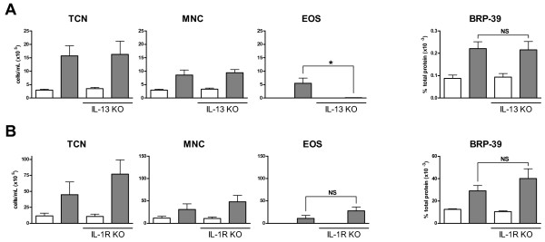
Figure 4.
HDM induced BRP-39 is IL-13 and IL-1 independent. WT BALB/c and IL-13 KO mice were saline (white bars) or HDM (grey bars) exposed for 10 days. (A) Data show total cell numbers (TCN), mononuclear cells (MNC), and eosinophils (EOS) in bronchoalveolar lavage fluid as well as BRP-39 expression in lung homogenates. (B) The same readouts are shown for IL-1R1 KO mice receiving HDM exposure. n = 5-10, data shown in B are representative of 2 separate experiments, * indicate P < 0.05.

