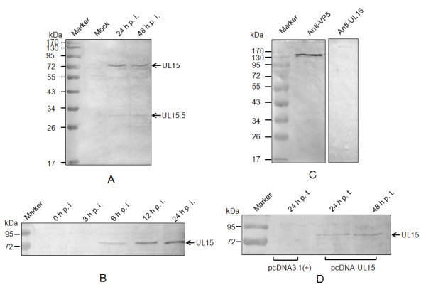
Figure 6.

Western blotting analysis of UL15 in infected cells and purified DEV virion. (A) Mock or DEV-infected cells were harvested at the indicated time post-infection, separated by SDS-PAGE and analyzed by western blotting using antiserum to UL15. The arrows at the right indicate UL15 and UL15.5. (B) Time-course of UL54 accumulation during DEV replication. (C) The purified DEV virion was separated by SDS-PAGE, blotted and immunodetected using UL15 antiserum, and McAb AB5 against DEV VP5 as a positive control. (D) pcDNA-UL15 or pcDNA3.1 (+)-transfected cells were harvested at the indicated time post transfection, and analyzed by western blotting using antiserum to UL15. The positions of molecular mass markers (in kDa) are indicated at the left.
