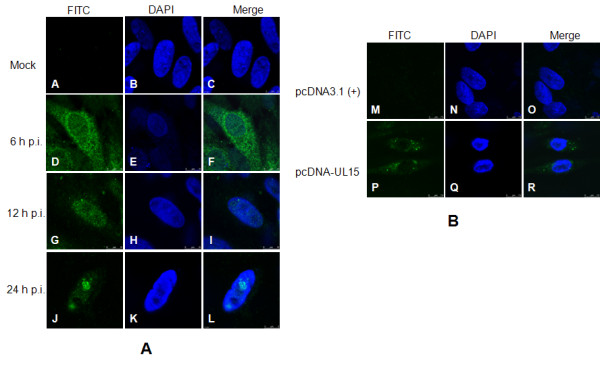
Figure 7.

Subcellular localization of DEV UL15 and/or UL15.5. CEF cells were seeded onto Fluorodishes (WPI, Sarasota, FL, USA) and were infected with DEV or transfected with pcDNA-UL15. At the indicated time post-infection, cells were fixed with 4% paraformaldehyde (Sigma-Aldrich), followed by permeabilizing with 0.5% Trixon X-100. Immunofluorescence analysis was performed by laser scanning confocal microscopy using the UL15 antiserum and FITC-conjugated secondary antibodies. Nuclear DNA was stained in all cases with DAPI. Columns in the up-down direction show the individual FITC (left) and DAPI (middle) images. Merged visualization of these the images are shown in the right-hand column.
