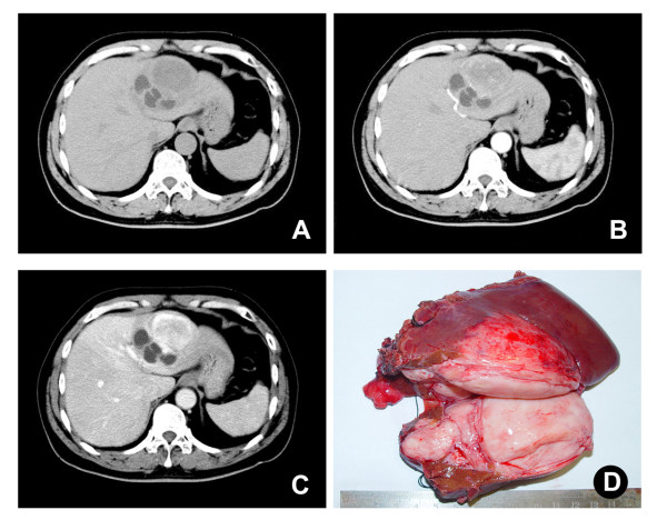
Figure 1.
Single mass in the left hepatic lobe. (A) CT scan demonstrated a mass (9.0 × 6.2cm) in left hepatic lobe. (B) and (C) Contrast enhancement in arterial and portal phases was found in the mass. (D) Grossly, a large, gray-white, lobulated, well-circumscribed, partially encapsulated mass was removed after hepatoectomy.

