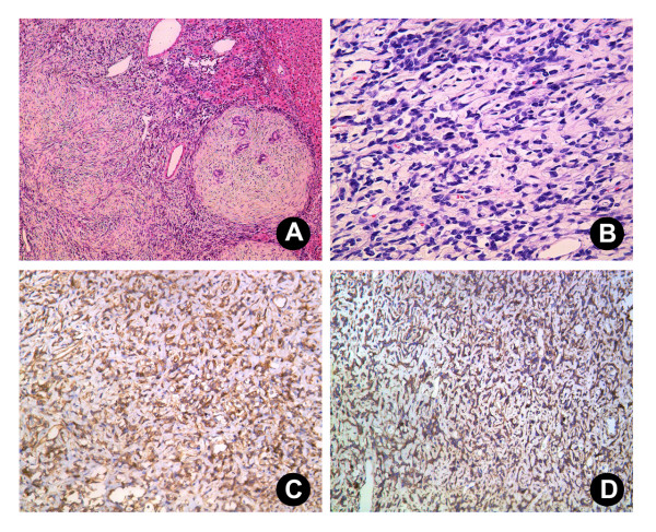
Figure 2.
Histological section of the SFT. (A) and (B) The tumor was composed principally of spindle cells arranged in short, ill-defined fascicles in some zones and randomly in others (A; H & E 10×; B; 40×). Immunohistochemically, the tumor cells were strongly positive for CD34 (C; 20×) and CD99 (D; 20×).

