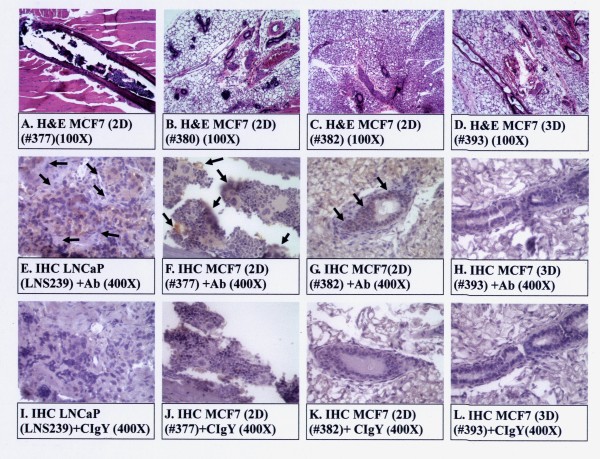
Figure 10.
Histology and immunohistochemistry (IHC) of MCF7 tumors grown in SCID mice. Histology of tumors derived from pooled 2D and 3D clones are shown in Panels A-D. IHC of tumors from pooled 2D and 3D clones are shown in Panels F-H and J-L. Tumors from the clone LNS239 of LNCaP cells subcutaneously injected to nude mice [3,10,32] was used as a positive control for IHC (panels E & I). The anti-human METCAM/MUC18 antibody (+Ab) was added to IHC in Panels E-H. Arrows show the positively stained cells by the anti-human METCAM/MUC18 antibody in the tumors derived from METCAM/MUC18-expressing MCF7 clones/cells (the pooled 2D clone). The control isotype antibody (CIgY) was added to the IHC in Panels I-L, as negative controls. Tumor sections from the tumors derived from the vector control 3D clone (Panels H&L) were also used as negative controls.

