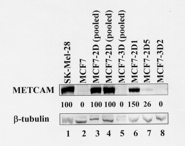
Figure 2.
HuMETCAM/MUC18 expression in various G418R clones of MCF7 cells. 5 μg proteins of each lysate were loaded in each lane except lane 4, in which 10 μg protein were loaded in Western blot analysis was used to determine the expression of METCAM/MUC18 in various clones/cell lines. Cell lysate from a human melanoma cell line, SK-Mel-18, was used as a positive control (lane 1) and that from the parental human breast cancer cell line MCF7 as a negative control (lane 2). METCAM/MUC18 expressions in cell lysates from five different MCF7 G418R clones (pooled 2D, pooled 3D, 2D1, 2D5, and 3D2) are shown in lanes 3-8. The number under lane indicates the relative level of METCAMMUC18 of each clone, assuming that in SK-Mel-28 as 100%. β-tubulin was used as the loading control.

