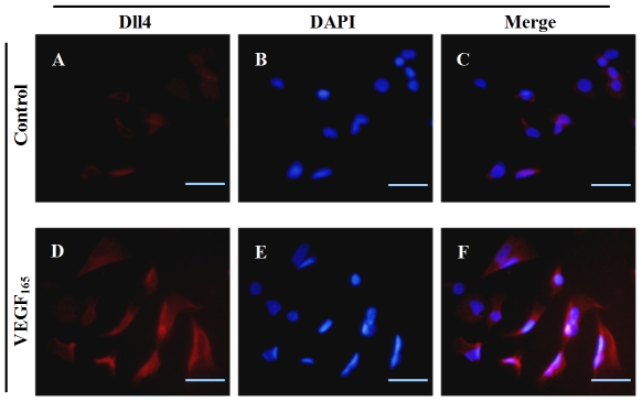
Figure 3. Representative immunofluorescence images of Dll4 in RF/6A cells stimulated by VEGF165.
CY3 (red)-conjugated secondary antibody represented Dll4 expression (A and D), DAPI (blue)-stained nuclei (B and E). (C) Merged image of A and B. (F) Merged image of D and E. The results clearly showed that Dll4 expression was induced by VEGF165 (D–F) compared with the PBS-treated control group (A–C). Bar = 50 µm.

