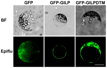
Figure 2. Plasma membrane localization of AtGILP.
Plasmids expressing GFP (left panel), GFP-AtGILP (middle panel), or GFP-AtGILPΔTM (right panel) were transfected into Arabidopsis mesophyll protoplasts. Fluorescent images were taken at 12–16 h after transfection. BF and Epiflu indicate bright field and epifluorescence, respectively. The scale bar is 20 µm. Results shown are representative of three independent experiments.

