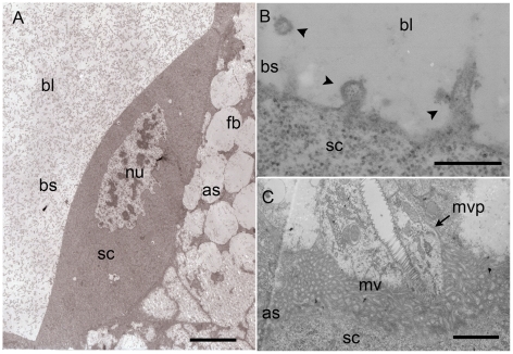
Figure 5. TEM micrographs of the serosa cells 48 h after oviposition.
A. Serosa cells (sc) are highly polarized with separate apical and basal membrane. B. The basal side (bs), facing the blastocoel (bl) shows several exocytosis vesicles (ve) (arrowheads in B). C. The apical side (as), facing the parasitized aphid tissue, shows microvilli (mv) and microvilli projections (mvp) in the aphid tissue. Scale bars = 10 µm in A, 1 µm in B and 3 µm in C. Abbreviations: as, apical side; bl, blastocoel; bs, basal side; fb, fat body; mvp, microvilli projections; mv, microvilli; nu, nucleus; sc, serosa cells.

