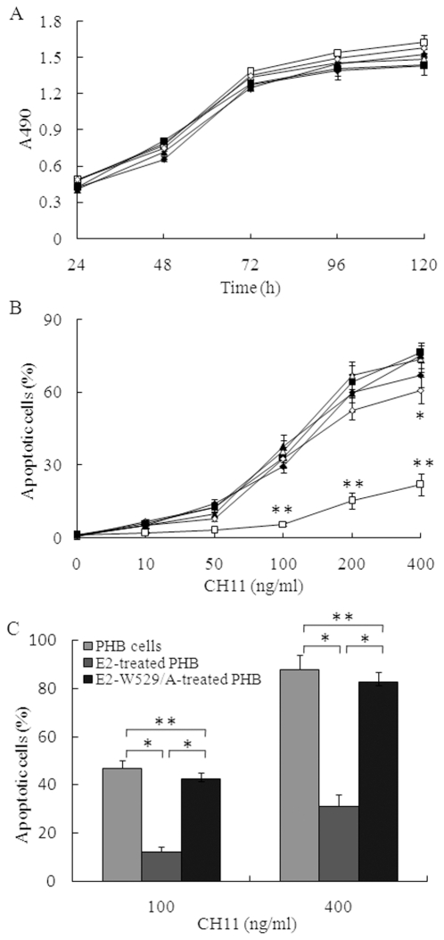
Figure 5. E2 blocks Raji cells apoptosis induced by anti-Fas antibody.
(A). Raji cells or CD81-silenced Raji cells were placed in 96-well plates coated with or without HCV E2 protein, cell viability was measured by MTS assay at various time courses. Data represent the means ± standard deviations of triplicate determinations. The treatments of the cells were: Raji cells cultured in 96 wells without coating with HCV E2 protein (open triangles), CD81 silenced Raji cells cultured in 96 wells without coating with HCV E2 protein (filled triangles), E2-treated Raji cells (open squares), E2-W529/A-treated Raji cells (filled squares), E2-treated CD81 silenced Raji cells (open diamonds), E2-W529/A-treated CD81 silenced Raji cells (filled diamonds). (B). Raji cells or CD81-silenced Raji cells were cultured in 96-well plates coated with or without HCV E2 protein for 24 h, and then incubated with CH11 at various concentrations for 5 h. Apoptotic cells were measured by Hoechst 33342 staining. Data points represent the means ± standard deviations of triplicate determinations. The treatments of the cells were described above. Student's t test was used to determine the statistical significance. Double asterisks, p<0.001 relative to other cell-treatment combinations. Asterisk, p<0.05 relative to the CD81 silenced Raji cells without treatment with E2 protein. (C). PHB cells were cultured in HCV E2 protein pre-coated 96-well plates for 24 h, and then incubated with CH11 at concentrations of 100 or 400 ng/ml. Apoptotic cells were measured after 5 h. Double asterisks, p>0.05. Asterisk, p<0.001.

