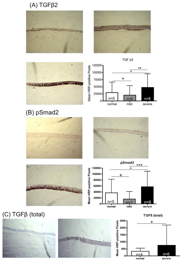
FIGURE 1.
Immunohistochemistry for transforming growth factor-β (TGFβ) signaling pathway markers. TGFβ2, pSmad2, and TGFβ (total) were assessed in normal human corneas and corneas with keratoconus. Horseradish peroxidase (HRP)-positive signal was quantified as described in Methods. Error bars indicate standard deviation. *P < .05; **P < .01; ***P < .001; +P ≥ .05; Mann–Whitney U test. Original magnification ×200. (A and B): Immunohistochemistry for TGFβ signaling pathway markers (TGFβ2 and phosphorylated Smad2 [pSmad2]) in (Top left) normal human corneas and (Top right) in patients with mild and (Bottom left) severe keratoconus. (Bottom right) Signal quantification. (C): Immunohistochemistry results for TGFβ (total) in (Left) normal human corneas and (Middle) patients with severe keratoconus. (Right) Signal quantification.

