FIGURE 2.

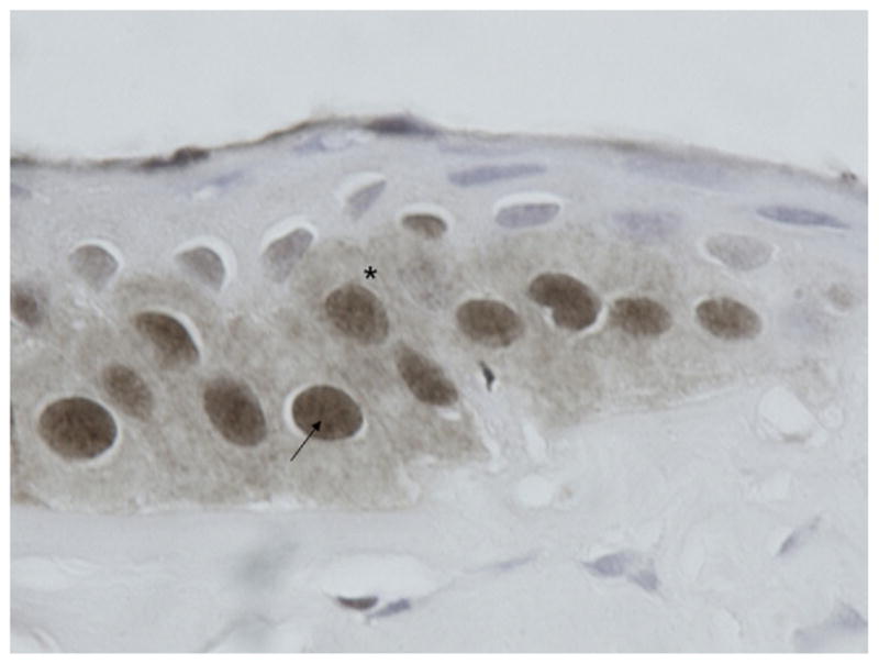
Photomicrograph showing immunohistochemistry results for phosphorylated Smad2 (pSmad2). Enlarged detail of keratoconus epithelium (×400 magnification) stained for pSmad2, showing a (*) perinuclear and (arrow) nuclear staining pattern.

Photomicrograph showing immunohistochemistry results for phosphorylated Smad2 (pSmad2). Enlarged detail of keratoconus epithelium (×400 magnification) stained for pSmad2, showing a (*) perinuclear and (arrow) nuclear staining pattern.