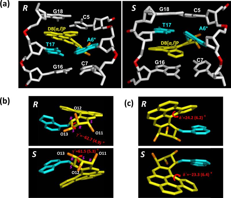
Figure 2.
(a) Views into the minor groove of the 14R (+)- and 14S (–)-trans-anti-DB[a,l]P-N6-dA adduct structures. These are the best representative structures for the 3.0 – 30.0 ns range of the MD simulations (See Methods). The central 3-mers are shown. (b) View into the minor groove side of the DB[a,l]P-modified adenine (c) View into the major groove side of the DB[a,l]P-modified adenine. The near mirror image nature of the stereoisomer pair is displayed; panel (b) emphasizes the fjord region twist and (c) emphasizes the benzylic ring pucker. For the DB[a,l]P moiety, the carbons are yellow and oxygen atoms are orange, the damaged bases are cyan. The DNA duplexes are white, except for the phosphorus atoms, which are red. Hydrogen atoms and pendant phosphate oxygen atoms are not shown for clarity. See also Movies S1 and S2, Supporting Information.

