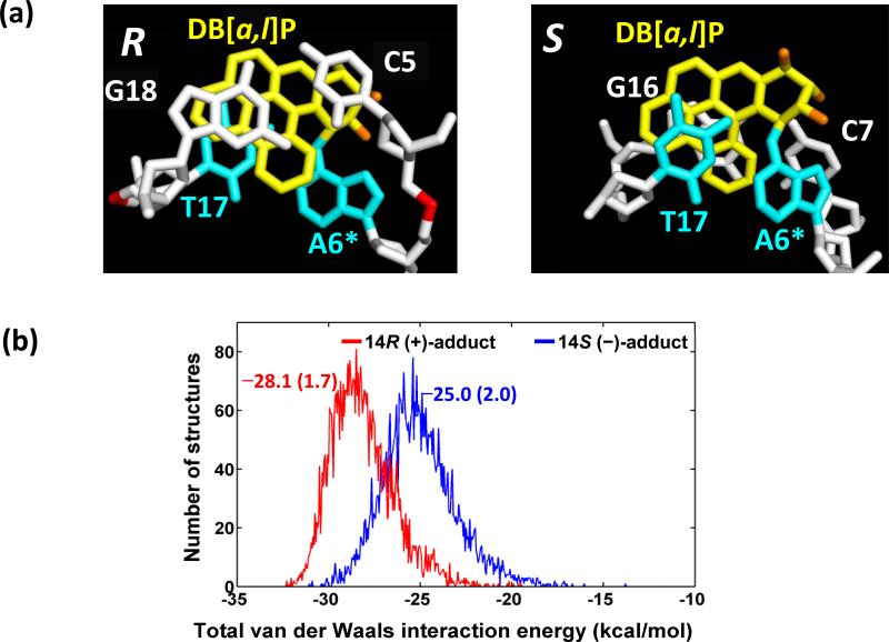
Figure 6.
(a) Views looking down the helix axis of the intercalation pockets. Note the substantial overlap between C5 and the DB[a,l]P rings in the 14R (+)-isomer, while the analogous C7 in the 14S (–)-isomer is poorly stacked. The structures shown are the best representative structures from the 3.0 – 30.0 ns MD simulations. Color schemes are the same as Figure 2. (b), population distribution of total van der Waals interaction energy between the DB[a,l]P aromatic rings and the intercalation pocket. Ensemble average values and standard deviation (in parentheses) (kcal/mol) are given.

