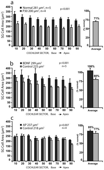
Figure 3.
a. Cross-sectional areas of the somata of spiral ganglion (SG) neurons are shown for 9 cochlear sectors representing 10% intervals from base to apex in normal controls and in neonatally deafened animals examined at 30 days of age (age when other animals were implanted). b. Cell size data are shown for neonatally deafened animals examined after 10 weeks of intracochlear BDNF infusion. Significantly larger cell size was documented in the BDNF-treated cochleae (black bars) as compared to the contralateral side (gray bars) in the same animals. After BDNF treatment, the ganglion cells measured about 300 μm2, which was equivalent to normal adult size and 33% larger than cells on the other side, which measured about 225 μm2. c. Data for control deafened animals after 10 weeks of infusion of artificial perilymph (AP). A modest, but significant difference in cell size is seen with larger cells in the AP cochleae than on the contralateral side. Comparison of the two data sets shows that cells in the BDNF-treated ears are markedly larger than those in all 3 other groups (cochleae contralateral to the BDNF-treated ears; AP cochleae, cochleae contralateral to the AP ears).

