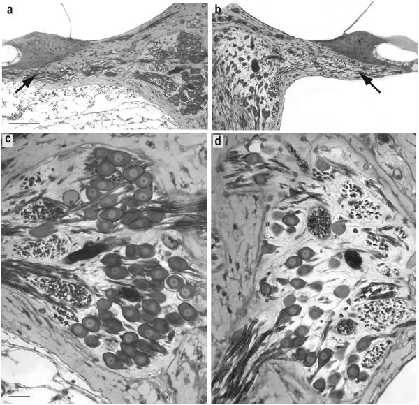
Figure 6.
Light microscopic images of histological sections from a neonatally deafened animal (K217) illustrating the marked neurotrophic effects of 10 weeks of unilateral intraocochlear BDNF infusion. The 40-50% cochlear sector of the left BDNF-treated cochlea (a,c) and the paired region from the contralateral deafened control cochleae (b,d) show a higher density of SG cells in Rosenthal's canal and a greater number radial nerve fibers within the osseous spiral lamina (arrows) after BDNF infusion. The fibrous tissue reaction to the implanted electrode is evident in the scala tympani of the left ear. The higher magnification images in c and d illustrate the quality of preservation and staining of the tissue after osmium post-fixation and the resolution of images used for morphometric analyses in the study. The higher density and size of SG perikarya in the BDNF-treated cochlea as compared to neurons on the opposite side are evident. (Scale bar in a = 50 μm; Scale bar in c = 25 μm).

