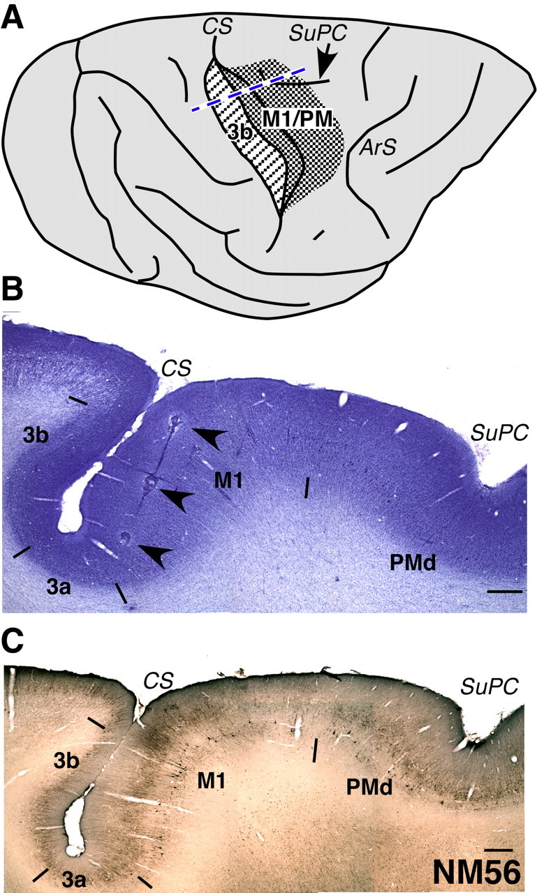
Figure 1.

A, A drawing of the dorsolateral view of a macaque monkey brain showing portions of the motor cortex (dense stippling) and the somatosensory area 3b (striped stippling) that were mapped. The densely stippled area included parts of both M1 and the premotor cortex (PM). The central sulcus has been opened for visualization in its depth. The blue dashed line shows the approximate location from where the sections shown in B and C were taken. ArS, Arcuate sulcus; CS, central sulcus; SuPC, superior precentral dimple. The rostral is to the right, and medial is to the top. B, A section of the brain from the normal monkey, NM56 stained for Nissl substance. The section was taken from the approximate location shown by the dashed line in A. The brain was cut in a plane perpendicular to the central sulcus (see Materials and Methods). An electrode track marked by electrolytic lesions is clearly visible (arrowheads). Borders of the somatosensory areas 3b, 3a, M1, and PMd are marked by short lines. The borders were determined by visualization in adjacent sections stained for Nissl, SMI-32 immunostaining (see C), acetylcholine esterase activity, and cytochrome oxidase activity. C, A section of the brain through the central sulcus stained by SMI-32 immunohistochemistry. The border between M1 and PMd is visible in such sections. In B and C, rostral is to the right. Scale bar, 1 mm.
