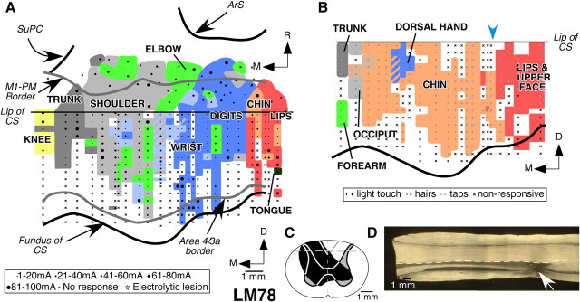
Figure 4.
Organization of the motor and somatosensory cortex in monkey LM78 with lesion of the dorsal columns on the left side. A, Organization of the motor cortex in a view similar to that shown in Figure 2. Note that the topography of the movement map is normal-like (compare Fig. 2). The ventralmost part of the motor cortex was partially mapped in this monkey (see Results). B, Somatotopy in area 3b of monkey LM78. The chin inputs (pink) expand medially into the deafferented hand area. Responses to touch on the hand were seen only at a few recording sites (blue). The estimated location of the prelesion hand–face border is marked by the blue arrowhead (see Results). For other conventions, see legend to Figure 3. C, Reconstruction of the spinal cord in a coronal plane in the region of the lesion, which was made on the left side. The extent of the damage is marked in black. The gray matter through the lesion and the border between the fasciculus cuneatus and fasciculus gracilis is shown for reference as mirror image of the right side. The dashed line marks the location from which the section shown in D was taken. D, A dark-field photomicrograph of a horizontal section of the spinal cord showing the lesion site (arrow). Rostral is to the left of the figure, and the left side of the spinal cord is toward the bottom. For abbreviations, see legends to Figures 1 and 2.

