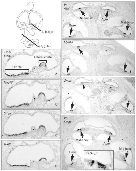
Fig. 3.
Expression patterns of Smpx and Tekt2 in the developing inner ear. Expression patterns of Smpx and Tekt2 in the developing inner ear analyzed with in situ hybridization at E15.5 (a-d), P1 (e-g), and P5 (h, i). Lines in the inner ear diagram show the levels of sections. Atoh1 (a, e) and Myo15 (b, f), well known markers for hair cells, were used to locate the hair cells in the inner ear tissues. At E15.5, expressions of Smpx and Tekt2 were found in the developing hair cells of the vestibular organs and continued up to P5 (a-d; data not shown), but were barely detected in the cochlea at this stage (data not shown). At P1, Atoh1 expression was found in the hair cells of all cochlear turns (e, arrows) and Myo15 expression was found in the base, mid-base, and mid-apex of the cochlea (f, arrows), but not yet in the apex (f, asterisk). Smpx expression was found in hair cells of basal and mid-basal turns (g, arrows) and weakly in the mid-apical turn (g, arrowhead). At P5, Smpx expression was detected in hair cells of all cochlear turns (h, i, arrows).

