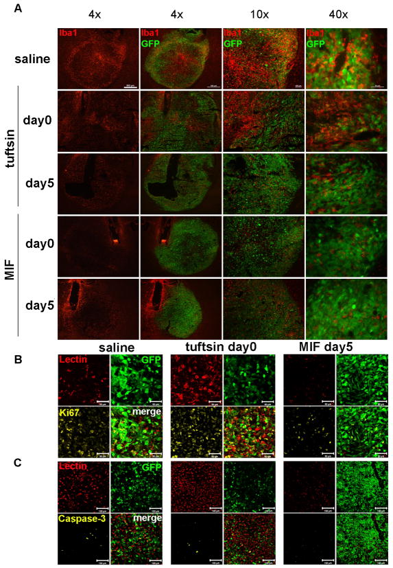
Figure 5. Tuftsin and MIF/TKP treatment affect glioma progression in vivo.
(A) Tumor sizes and MG/MP infiltration (Iba1+ cells) were evaluated 14 days after GL261-EGFP injection and infusion of 250μg/ml tuftsin (TF), MIF/TKP or saline (scale bar 500μm). The proliferation and apoptosis of tumor cells and GIM in saline group, D0 tuftsin and D5 MIF/TKP groups were quantified with (B) Ki67 (scale bar 50μm) and (C) activated caspase-3 (scale bar 100μm) immunofluorescent staining (n = 3).

