
Figure 2. Histological variants of massive hepatic necrosis (MHN).
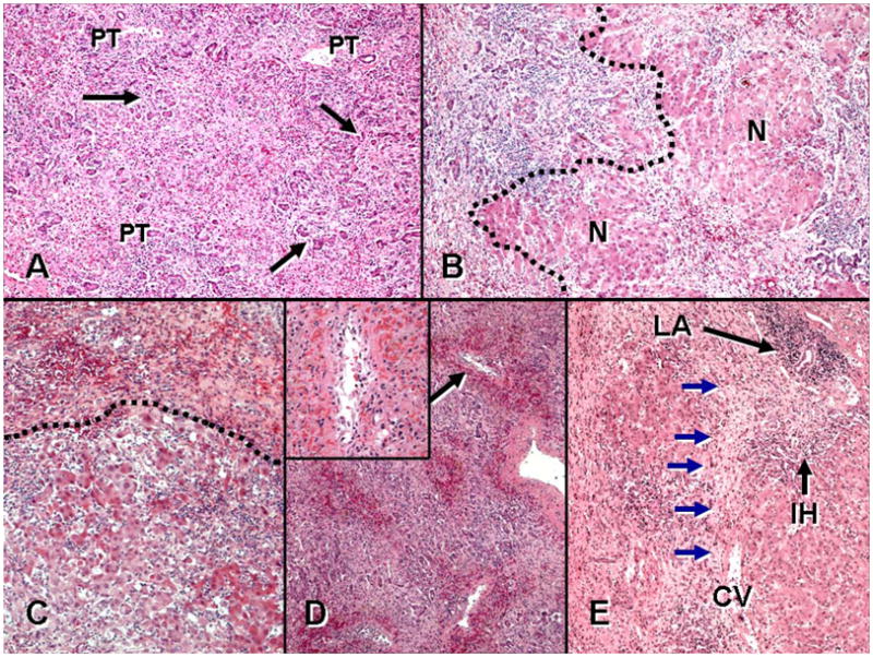
A. MHN type 1: “typical” MHN with panlobular necrosis and neocholangiolar proliferation (ductular reaction) (arrows) emerging from parenchyma near portal tracts (PT). B. MHN type 2: there is confluent necrosis in the field left of the hatched line, with early regenerative nodules (N), a pattern also referred to as ‘submassive necrosis’. C. MHN type 3: acute hepatitis with lobular disarray, inflammation and liver-cell damage is seen at bottom and is separated by the hatched line from more severe confluent necrosis above. D. MHN type 4: centrilobular hemorrhagic necrosis superimposed on the general features of massive necrosis is prominent at low power. Inset: one centrilobular region (arrow) shows lymphocytes and plasma cells around, infiltrating, and within the lumen of a central vein (‘central perivenulitis’), in association with hemorrhage and hepatocyte loss. E. MHN type 5: confluent necrosis is present superimposed on features of an underlying chronic hepatitis, including portal lymphoid aggregates (LA) and interface hepatitis (IH). Note confluent bridging hepatic necrosis (blue arrows) extending between central vein (CV) and portal tract at upper right. Multilobular necrosis is also present at the left of the field. (A-E: Hematoxylin and eosin stain, × 40. Inset to D: Hematoxylin and eosin stain, × 200.)