
Figure 3. Central perivenulitits.
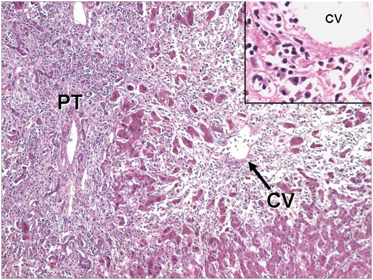
This section from an explanted liver shows severe hepatitis with extensive inflammation extending from the portal tract (PT) into the lobule, with marked loss of hepatocytes. Note the prominent hepatocyte loss and inflammation around the central vein (CV), “central perivenulitis.” Inset: Higher magnification of central perivenulitis highlighted by plasma cells surrounding and infiltrating the vein wall, associated with marked hepatocyte destruction. (Hematoxylin and eosin stain).