
Figure 2.
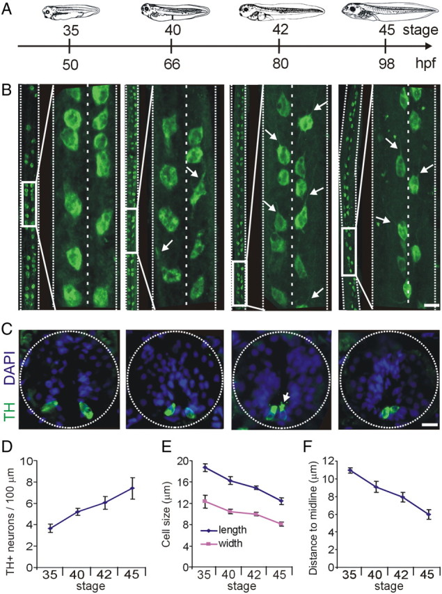
Development of spinal cord dopaminergic neurons. A, Developmental timeline as in Figure 1. B, Whole-mount projections of spinal cords stained for TH immunoreactivity. At each stage, large midbody segments of spinal cord are shown on the left side, and an inset expansion is shown on the right. Projections are shown ventral side and rostral end up. Arrows on the stages 40, 42, and 45 panels point to axons extending rostrally. C, Cross sections through the spinal cord at different stages of development. Dopaminergic spinal neurons are located on the ventral side of the spinal cord. The arrow points to microvilli and cilia extending into the central canal. D, Quantification of the number of neurons per 100 μm of spinal cord at different stages of development. E, Length and width measurements of dopaminergic spinal neurons. F, Quantification of the distance to the midline. Scale bars, 15 μm. Values are mean ± SEM for n ≥ 4 larvae per stage.