
Figure 3.
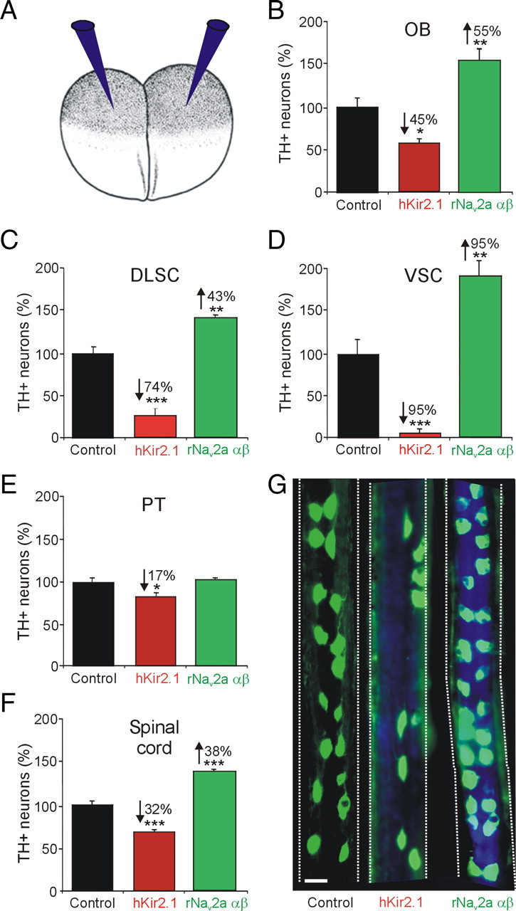
Dopaminergic specification is activity dependent. A, Embryos at the two-cell stage were injected bilaterally with either hKir2.1 or rNav2a αβ mRNA along with Cascade Blue tracer, to decrease or increase calcium spike activity, respectively (Borodinsky et al., 2004; Dulcis and Spitzer, 2008). B–F, Number of TH+ neurons (percentage) in activity-manipulated embryos in the OB (B), DLSC (C), VSC (D), PT (E), and spinal cord (F). G, Spinal cord whole mounts from stage 42 larvae stained for TH immunoreactivity (in green) show the effect of channel misexpression. The tracer (in blue) is seen in spinal cords from activity-manipulated larvae. Numerical percentage change is indicated in cases of significant difference (n ≥ 6 larvae per stage; values are mean ± SEM): *p < 0.05, **p < 0.01, ***p < 0.001, comparing control values with hKir2.1 or rNav2a αβ. The Mann–Whitney U test was used to assess statistical significance. Scale bar, 25 μm.