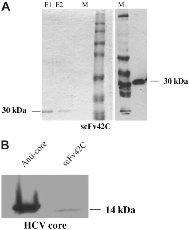
Fig. 3.
Purification of functional scFv42C and its binding to HCV core protein. (A) Detection of purified scFv42C. Coomassie blue staining of purified scFv42C separated in SDS-PAGE (left). The scFv42C antibody was purified using Ni-NTA agarose. E1: 1st elution; E2: 2nd elution. The predicted size of scFv is 30 kDa. Immunoblotting of purified scFv42C with α-his mAb (right). M: full-range rainbow molecular mass protein standard. (B) Binding of bacterially expressed and purified scFv42C fragment to HCV core protein. The 14 kDa of core protein (aa 1-115, Mikrogen) was separated in 12% SDS-PAGE and blotted onto membrane. Lane 1: detection with α-HCV-core antibody (C7–50, ABR) followed by α-mouse-HRP. Lane 2: detection with FLAG-tagged scFv42C, followed by α-FLAG mAb and α-mouse-HRP. [Color figure can be viewed in the online issue, which is available at www.interscience.wiley.com.]

