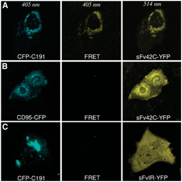
Fig. 6.
FRET analysis. (A) Specific interaction between scFv42C and HCV core protein. Huh-7 cells were cotransfected with CFP-C191 and scFv42C-YFP constructs. CFP was detected at a wavelength of 405 nm and YFP at 514 nm. The observed YFP-specific spectrum in perinuclear region using 405 nm excitation clearly reveals an assignment of the resonance energy from CFP-C191 to sFv42C-YFP (FRET, middle panel). (B, C) Negative controls of FRET. Huh-7 cells were cotransfected with CD95-CFP and sFv42C-YFP or CFP-C191 and scFvIRYFP. No YFP-specific spectrum was detected using 405 nm excitation (middle panel).

