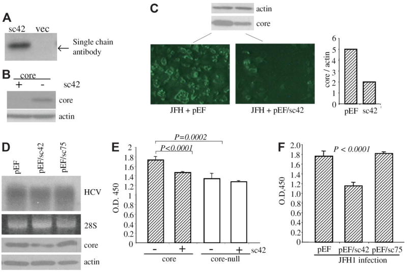
Fig. 7.
Effect of scFv42C on core protein expression, replication, and cell proliferation. (A) Intracellular expression of scFv42C. Huh-7 cells were transfected with pEF/sc42 or empty vector (vec) and harvested 2 days later. Expression of scFv42C was detected by Western blot using HisProbe. (B) Effect of scFv42C antibody on core protein level. Huh-7 cells were co-transfected with 0.5 μg of pZeo+/core and pEF/sc42 or empty vector. Core protein was analyzed by Western blot at day 2 after transfection. Actin was detected in parallel as loading controls. (C) Immune staining of core protein expressed from JFH1 replicon. Huh-7.5 cells were coelectroporated with 5 μg of replicon RNA and 2 μg pEF/sc42 plasmid or empty vector, and seeded in 24-well plates with cover slips. Cells were fixed 3 days later and stained with anti-core antibody followed by ultraviolet microscopy as described. 10 Cells seeded in six-well plates were subject to Western blot of core (10 μL lysate) and β-actin (2 μL lysate). The relative ratio of the core versus actin in Western blot was measured by densitometry (right panel). (D) Effect of scFv42C on HCV replication. Huh-7.5 cells were electroporated with 5 μg of JFH replicon RNA and continuously cultured under subconfluent condition for 76 days. Cells were then electroporated with 2 μg plasmid DNA encoding scFv42C (pEF/sc42), or nonrelevant scFv (pEF/sc75), or empty vector (pEF). Viral replication was examined by Northern hybridization. The 28S RNA served as loading controls. Core and β-actin expression was also shown. (E) Effect of scFv42C on cell proliferation. Huh-7 cells were cotransfected with pZeo +/core (or a core-null mutant as a control) (0.19 μg/six wells of a 96-well plate) and same amount of pEF/sc42 (or empty vector). Cell proliferation was measured by a modified MTT assay (cholecystokinin-8 assay) at day 1 after transfection. Core-null: core cDNA with a stop codon immediately downstream of the ATG codon. (F) Effect of single-chain antibody on cell proliferation in the context of HCV replication. HCV-infected Huh-7.5 cells as described in (D) were seeded in 96-well plates after electroporation with the plasmids indicated. Cell proliferation was measured 2 days later as described above. Data are presented as the mean ± standard deviation (n = 6). Statistical analysis was done by Student t test.

