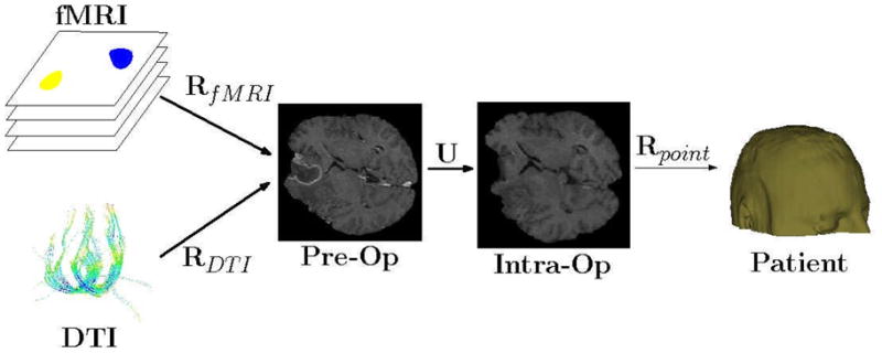
Figure 3.

Transformation cascade. Three steps are needed to map the functional images, fMRI and DTI, into the intra-operative physical space. First, rigid, or affine, transforms, RfMRI and RDTI are estimated to align the functional images with the pre-operative anatomical image. A rigid transform is generally adequate because the brain is still enclosed in the skull and assumed to move rigidly up until the start of the surgery. Secondly, a non-rigid transform U is estimated to align the anatomical pre-operative image with the anatomical intra-operative image. A non-rigid transform is required to capture the non-linear movements due to for instance brain-shift. A rigid transform Rpoint is estimated from homologous points extracted from the patient space and the intra-operative image. In some intra-operative MR scanners, for instance the GE Signa SP 0.5T which was in use at BWH until 2007, the physical tracking space and the image space were aligned and the estimation of Rpoint was unnecessary. Consequently, points X in the pre-operative functional space can be mapped to the patient (physical) space with the following transformation cascade: Rpoint (U(RfMRI(X))). This enables navigation of the functional images in a neuro-surgical navigation system.
