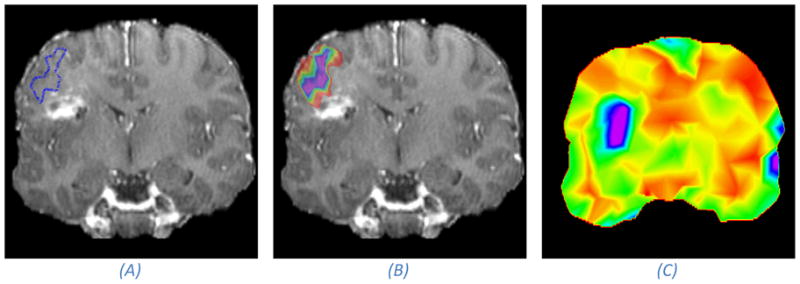
Figure 5.

Illustration of the effect of registration uncertainty on mapping of functional areas from the pre-operative to the intra-operative space. The anatomical image is a post-operative image, acting as a proxy for an intra-operative image, acquired from the same patient we show pre-operative images of in Figure 1. (A) A coronal MRI slice with the estimated boundary of a speech activated functional area after registration. (B) Takes into account the registration uncertainty, and rather than visualizing the point-estimate as in (A), it visualizes the marginal probability that a certain voxel is inside the functional area (purple denotes a probability of one which falls off normal to the iso-contours down to red which is close to a zero probability). If we assume that the hyper intense area is the site of the resection, then we can conclude with a small probability that the functional area was touched during surgery because the outer rim of the color map touches on the “resected” area. Image (C) shows an uncertainty map. The uncertainty is measured as the dispersion of the marginal posterior distribution over deformation parameters. We have colored high uncertainty with purple and low uncertainty with red. It can be seen that the registration uncertainty is higher close to the resection than away from the resection. It is important to keep in mind that a high uncertainty in the registration does not necessarily mean that the registration is inaccurate, but the chance that it is less accurate is higher.
