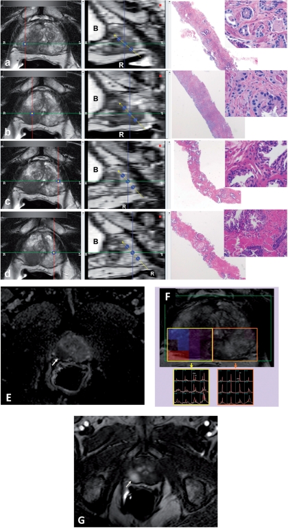
Figure 2.
The software used for MRI analysis at sextant specimen locations. Left column windows show multi-planar reconstructed images perpendicular to the biopsy core (single blue dot); the middle column windows show the sagittal views aligned with the tract of the biopsy core (triple blue dots with dashed yellow line) (a–d). T2W MRI findings of RML (a), RMM (b), LML (c) and LMM (d) peripheral zone biopsy core sites of which RML and RMM core sites show positive MRI findings for tumor, whereas LML and LMM core sites appear normal. The biopsy results for RML (a), RMM (b), LML (c) and LMM (d) are Gleason 4 + 4 (70%), Gleason 4 + 5 (50%), benign and benign, respectively. Right column windows show the corresponding hematoxylin/eosin stained biopsy images with 2× and 40× magnification. RML, right mid lateral; RMM, right mid medial; LML, left mid lateral; LMM, left mid medial; R, rectum; B, bladder. Multi-parametric MRI sequences (ADC maps of DW MRI (e), MRS (f) and DCE MRI (g)) localize the right sided tumor (arrows).

