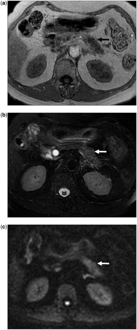
Figure 10.
(a) A 67-year-old man with typical non-contrast MRI features for PCa. Axial T1-weighted gradient recalled echo MR image at the level of the body of the pancreas showing the typical appearance for PCa. Tumor (arrow) is hypointense to the pancreas. (b) Axial fat-suppressed T2 fast spin echo MR image at the same level as (a) shows the same infiltrative mass as mildly hyperintense (arrow). The pancreas itself is not well visualized due to its relatively low signal. (c) Axial diffusion-weighted MR image (b-value 1000 s/mm2) showing heterogeneous increased signal in the body of the pancreas consistent with a tumor (arrow).

