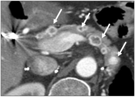
Figure 12.
A 46-year-old woman with a history of renal cell cancer. Axial contrast-enhanced CT image of a patient with a history of renal cell carcinoma showing the typical appearance of renal cell carcinoma metastases. In this case, multiple lesions (arrows) are present throughout pancreas, alluding to metastatic disease. The lesions all show central hypodensity with avid peripheral enhancement. However, there is no pancreatic ductal dilation.

