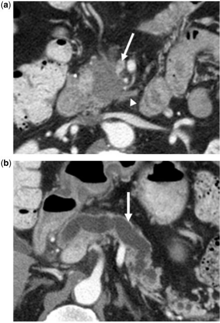
Figure 14.
(a) A 65-year-old man with a history of PCa. Axial contrast-enhanced CT image showing tumor presenting as an irregular hypodense mass in the pancreatic head. The tear-drop sign of the superior mesenteric vein (arrow) is consistent with vascular invasion. Perineural (arrowhead) invasion is likely present and involving the superior mesenteric plexus given tumor extension along the inferior pancreaticoduodenal artery. (b) Gross dilatation of the pancreatic duct (arrow) is also present and supportive of diagnosis of primary pancreatic cancer.

