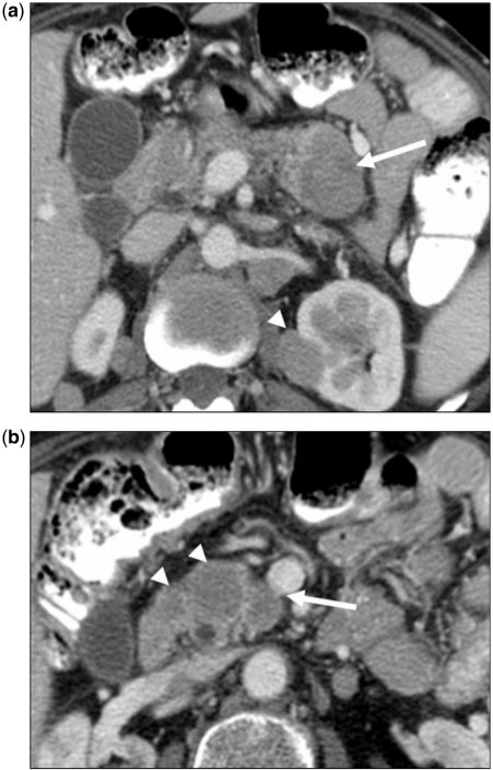
Figure 2.
(a) A 49-year-old woman with a history of cervical cancer. Axial contrast-enhanced CT image showing hypovascular metastasis (arrow) in the pancreatic tail. A second deposit is demonstrated in the left kidney (arrowhead). The pancreatic metastasis is hypoattenuating, similar to pancreatic adenocarcinoma (PCa). Unlike PCa, this metastasis is relatively well defined, and is not associated with pancreatic ductal dilatation or regional vascular invasion. (b) Contrast-enhanced CT image at a more inferior position in the pancreas showing additional lesions (arrowheads). Metastases were homogeneously hypodense to pancreatic parenchyma. The superior mesenteric vein is intact even though one lesion (arrow) lies just adjacent to it. Lack of vascular invasion favors metastatic disease over PCa. However, multifocal pancreatic involvement is the strongest factor supportive of metastatic disease.

