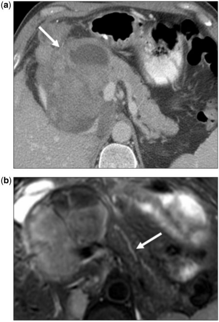
Figure 3.
(a) A 45-year-old man with a history of retroperitoneal sarcoma. Axial contrast-enhanced CT image of a patient with unclassified retroperitoneal sarcoma metastasizing to the pancreatic head. Large lesion (arrow) is heterogeneously hypodense to the rest of the pancreas, however does not cause pancreatic ductal dilation or occlusion of the superior mesenteric vein (arrowhead). (b) Axial fat-suppressed T2-weighted fast spin echo image of the same lesion showing minimally prominent caliber of the pancreatic duct (arrow). Imaging findings favor metastatic disease, as pancreatic adenocarcinoma of this size would be expected to cause significant biliary and pancreatic ductal dilatation.

