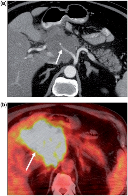
Figure 4.
(a) A 75-year-old woman with non-Hodgkin lymphoma. Axial contrast-enhanced CT image showing a hypodense mass in the pancreatic head. The common hepatic artery (arrow) is encased. Note there is no pancreatic ductal dilation. (b) Corresponding fusion positron emission tomography (PET)/CT image shows marked hypermetabolism in keeping with the primary diagnosis. However, such a finding is not specific, as even primary pancreatic adenocarcinoma would be expected to show increased metabolic activity.

