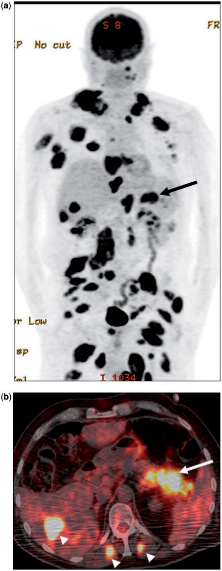
Figure 5.
(a) A 62-year-old man with known metastatic melanoma. Coronal PET image showing extensive deposits throughout the body. A large metastatic deposit was present in the pancreatic tail (arrow). (b) Corresponding fusion PET/CT image in the axial plane confirms the anatomic location of the lesion (arrow). Note the presence of extrapancreatic metastases (arrowheads) in the liver and bilateral erector spinae muscles. The latter are rarely seen in PCa, but a fairly frequent finding in metastatic melanoma.

