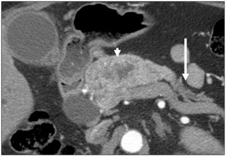
Figure 9.
A 55-year-old man with a history of renal cell carcinoma. Axial contrast-enhanced CT image showing the infiltrative pattern of a hypervascular metastasis (arrowhead) in the proximal pancreas. This metastasis was associatd with biliary obstruction (not shown), likely secondary to the relatively large size of the metastasis and its location. Biliary obstruction is more common with larger metastases. Overall, the frequency of associated biliary obstruction is less than that for primary pancreatic cancer. Note the atrophic distal pancreas with associated ductal dilatation (arrow), a sign of chronic obstruction.

