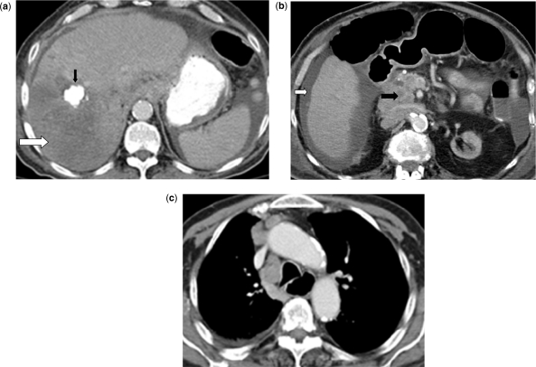
Figure 3.
Abdominal CT scan reveals a 13 × 10 cm recurrent hypodense liver mass that replaced most of the right lobe (a) (white arrow), a calcified nodular lesion in the right lobe due to prior chemoembolization (black arrow), ascites (b) (white arrow), metastatic adenopathy in the mesentery, retroperitoneum (b) (black arrow). Chest CT scan reveals metastatic mediastinal adenopathy (c).

