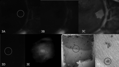
Figure 3.
Impression cytology of a punctate spot. (A) Clinical photograph of limbal area exhibits a hyperfluorescence spot, encircled. Linear limbal fluorescence provided fiducials. (B) Disappearance of the punctate spot after impression cytology. (C) After repeat instillation of fluorescein, a hyperfluorescent outline of the membrane remains. (D) Enlargement of the punctate spot encircled in (A) and the corresponding spot on the impression membrane, viewed with epifluorescence (E). The hyperfluorescent spot localized to a cell (circle) after rapid, air-dried staining of the membrane (F), which features less distinct cytoplasmic and nuclear borders than do the adjacent cells (G). Original magnification: (A–D) ×10; (E, F) ×100; (G) ×400.

