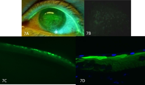
Figure 7.
Confocal microscopy of punctate spots. Clinical photograph of punctate spots immediately before corneal transplantation (A). Epifluorescence photograph of the corneal button demonstrates punctate stains. Preoperative fluorescein was still present in the tissue (B). Punctate staining of cornea in cross section (C). After DAPI staining, punctate stains appeared in the second cell layer (D). Original magnification: (A) none; (B) ×100; (C) ×100; (D) ×600.

