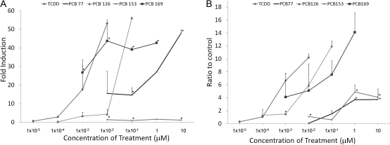
FIG. 3.
Different DLCs induce Cyp1a1 expression in proportion to their TEF. (A) Hepa-1 cells were treated for 8 h with the indicated concentrations. After treatment, RNA was extracted and used for QRT-PCR determination of the amount of Cyp1a1 mRNA relative to β-actin. (B) Hepa-1 cells were treated with the indicated concentrations of TCDD, DMSO, or PCBs for 90 min. Cells were then fixed, and ChIP was performed for AHR. Error bars represent SE. An * indicates statistical significance when compared with the similar TEQ of TCDD (p value < 0.05).

