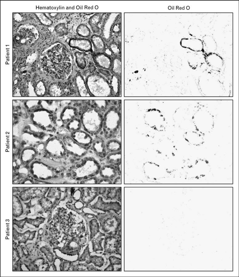
Figure 1. Lipid accumulation in human kidney samples visualized by Oil Red O staining.
Kidney surgical specimens were obtained from patients undergoing radical nephrectomy for renal cell carcinoma. Normal kidney cortex samples were dissected by an experienced pathologist, away from the tumor. The samples were frozen, sectioned, and stained with hematoxylin and Oil Red O to visualize the distribution of lipids within renal structures. Left panels are representative images from three different patients (original color images are shown here in gray scale). For each image, a computer-based color deconvolution algorithm was used to separately visualize Oil Red O staining in the red channel (right panels). In these examples, lipid deposits are localized mostly within tubular epithelial cells in patients 1 and 2, but are not detectable in patient 3.

