Figure 2.
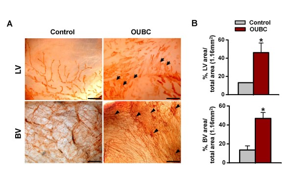
OUBC displays markedly increased number of lymphatic and blood vessels in the peritoneal surface of bladder wall. PBS (Control) or 1 × 10⁶ MBT-2 cells (OUBC) were injected into the urinary bladders of 8-10-week old female C3H mice. 4 weeks after PBS or the tumor cell injection, bladder was whole-mounted and immunostained for LYVE-1 and PECAM-1. (A) LYVE-1+ LVs and PECAM-1+ BVs in bladder surface were visualized with DAB staining (brown). Scale bars, 200 μm. Arrows indicate profound and tortuous lymphangiogenesis and arrowheads indicate robust increase of BV networks. (B) Comparison of lymphatic vessel densities (LVD) and BVD per total area (1.16 mm2) of bladder. Graph shows mean ± SD; n = 5 for each group.*p < 0.05 versus Control.

