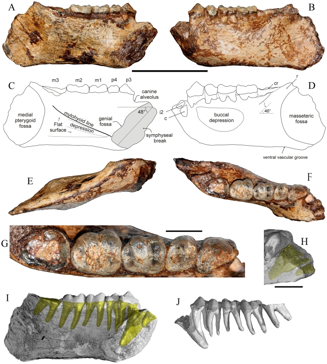
Figure 2. MFI-K171, holotype mandible of Khoratpithecus ayeyarwadyensis n. sp.
A: lingual view. B: buccal view. C: interpretive drawing of the lingual view. D: interpretive drawing of the buccal view. E: ventral view. F: occlusal view. G: enlarged occlusal view of the tooth row. H: Detailed view of the anterior region displaying alveoli shadows. Scale bars: 5 cm for A–F and I–J and 1 cm for G–H. Abbreviations: cr, crown remnant; r, ramus. I: lingual view displaying roots shadows for P3-M3 and alveoli shadows for incisors and the canine. J: dental row with virtually extracted roots in buccal view.

