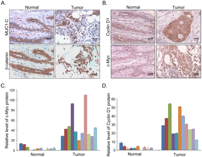
Figure 6. MUC1-C is associated with increased β-catenin nuclear localization and activity in clinical breast carcinomas.
(A) Representative images of IHC staining of MUC1-C and β-catenin in sections from human breast cancer tissue and normal mammary tissues. (B) Representative images of IHC staining of Cyclin D1 and c-Myc in sections from human breast cancer tissues and normal mammary tissues. Intensity analysis of c-Myc (C) and Cyclin D1 (D) in sections from 11 human breast cancer tissue samples and 11 normal mammary tissue samples. The data represented as relative fold of intensity of IHC staining in graphs were measured using Image-Pro Plus software; scale bars, 30 µm.

