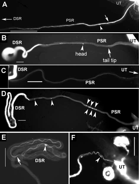
FIG. 2.
Parallel formation of flagella during SR entry and exit. Still images from frozen samples are shown. A–C) Images of sperm entry movement were obtained 15 min to 1 h after mating. A) Sperm entered the PSR in a parallel formation consisting of multiple sperm tails twisting around each other like a bundle. This tail bundle has entered halfway (0.5 mm) into the PSR. One head (arrowhead) and one flagellar loop (arrow), representing sperm with atypical flagellar configurations, were found in the mix of the tail bundle in A (Fig. 3F and Supplemental Movie 9). An explanation for these atypical sperm is given in Figure 8. B) Two sperm exhibit tail-leading PSR entry movement: One just entered the PSR, and the other nearly entered the DSR. The flagella of these sperm were in extended linear shape (a small fold was caused by unfolding the naturally folded PSR). C) Sperm moved across the PSR lumen without flagellar folds. D and F) Images of sperm exit movement were obtained 24 h after mating, and sperm exhibited head-leading movement (arrowheads indicate sperm heads). E) A sperm inside in the DSR is folded (arrow indicates, tail tip). Bar = 0.1 mm.

