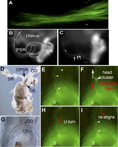
FIG. 3.
Leading tails and lagging heads. The dynamic process of PSR entry is shown here with video snap views of live samples. A) Multiple sperm tails enter the PSR in parallel formation like a bundle; the sperm are labeled with both head and tail GFP (Supplemental Movie 6). B and C) The sperm (labeled with head GFP only) pass through the convoluted PSR tubule (line trace in B) and enter the folded DSR on the right, which already contained a lot of sperm, as indicated by the bright fluorescence (Supplemental Movies 7 and 8). Arrows in C point to a cluster of three sperm heads that moved in one overlapping array consisting of eight sperm, which was then followed by a second array consisting of five sperm. The array is defined by having head-to-head spacing shorter than one sperm length of 2 mm (Supplemental Table S1). All of these sperm went through the PSR without to-and-fro motion and without turning or changing of the direction of movement. The time was 45 min after mating. D) Image of the uterus shows the relative locations of the oviduct (OD), ventral SR, and dorsal spermathecae (S). Arrows point to the openings of SR and spermathecae (OPSR and OS, respectively). G) Image at the uterus-oviduct junction provides spatial orientation for Supplemental Movie 9 (E, F, H, and I). Notice that the spermathecae are folded back onto the dorsal surface of the uterus. The dashed circle represents the location of the sperm focus, from which sperm initiate PSR entry movement in Supplemental Movie 9. The dashed arrow points to the direction of moving parallel sperm array. E, F, H, and I) Sequential snap views from Supplemental Movie 9 (time index, 1.375, 1.750, 3.375, and 4.375 sec). When multiple sperm flagella align into a parallel bundle, this appears as a GFP trail that leads upward toward the OPSR. E) Two sperm heads are on the GFP trail, and a head cluster is just about to be pulled out of the circular sperm focus in the uterus. F) The head cluster is pulled out and moving upward on the GFP trail. The head cluster contained one atypical sperm (red arrowhead), which is identifiable from its opposite moving direction and separation from the head cluster. The white arrow points to the upward-moving direction of most sperm, whereas the red arrow points to the downward-moving direction of the atypical sperm. H and I) The atypical sperm head undergoes a U-turn (H), and it rejoins the majority of sperm on the GFP trail (I). Original magnifications ×400 (A), ×20 (B, C), ×10 (D), ×40 (E–I).

